


Ultrasound Guided Diagnostic Fluid Tap ( Procedure Charges Only)
ULTRASOUND
Ultrasound Guided Diagnostic Fluid Tap ( Procedure Charges Only)
Also Known As:
Ultrasound-Guided Diagnostic Tap
USG-Guided Diagnostic Fluid Sampling
USG-Guided Fluid Aspiration
USG Pleural tapping
Usg Ascitic fluid tapping
₹4,000
About This Test
Ultrasound-guided tapping, also known as aspiration, is a minimally invasive procedure used to remove fluid from body cavities such as the abdomen (ascites), pleura (pleural effusion), or joints. Real-time ultrasound guidance ensures accurate needle placement, minimizes complications, and avoids injury to adjacent organs or vessels. The procedure can be diagnostic, allowing fluid analysis for infection, malignancy, or other pathologies, or therapeutic, providing symptom relief from fluid accumulation. Ultrasound guidance improves safety, efficiency, and patient comfort. It is widely used in emergency, critical care, and outpatient settings, making it an essential tool in clinical practice.
At a Glance
Home Collection
Not Available
Report TAT
1 Day
Category
ULTRASOUND
Preparation / Prerequisite
Bring latest BT, CT & PT report. Old record to be shown & assessed by radiologist whether procedure is possible & required or not. If any non invasive procedure is required before invasive procedure, please discuss with referring doctor.
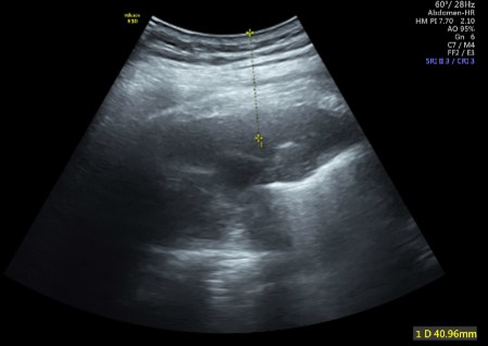
Reference Image
Frequently Asked Questions
1. What is ultrasound-guided diagnostic fluid tapping?
Ans. It is a small invasive procedure where fluid is removed from the body using a thin needle.
Q
2. Why is this procedure done?
Ans. It is done to
Find out why fluid has collected (for eg . Abdomen (ascites), Chest (pleural fluid), Joint & Around organs or swellings),
Check for infection, cancer, or other disease
Q
3. Is it safe?
Ans . Yes completely safe & No radiation is used.
Q
4. Is any preparation needed?
Ans. Old record to be shown & assessed by radiologist whether procedure is possible & required or not. You cant eat and drink normally.
Q
5. How long does the scan take?
Ans. It usually takes 15-20 minutes.
Q
6. Is appointment required ?
Ans. Yes, prior appoinment is required .
Q
7. Is an attendant required ?
Ans . It is mandatory to bring an attendant with you .
Q
8. Is there any need to bring previous records ?
Ans. Yes , bring relevant old records.
TAT Policy
The Turnaround Time (TAT) depends on the following factors:
- Registration date and time
- Type of scan or test
- Scan time or sample collection time
In uncommon circumstances, TAT may be delayed due to test complexity or reasons beyond our control. You will be informed of the estimated TAT at the time of registration.