


What is PET-CT
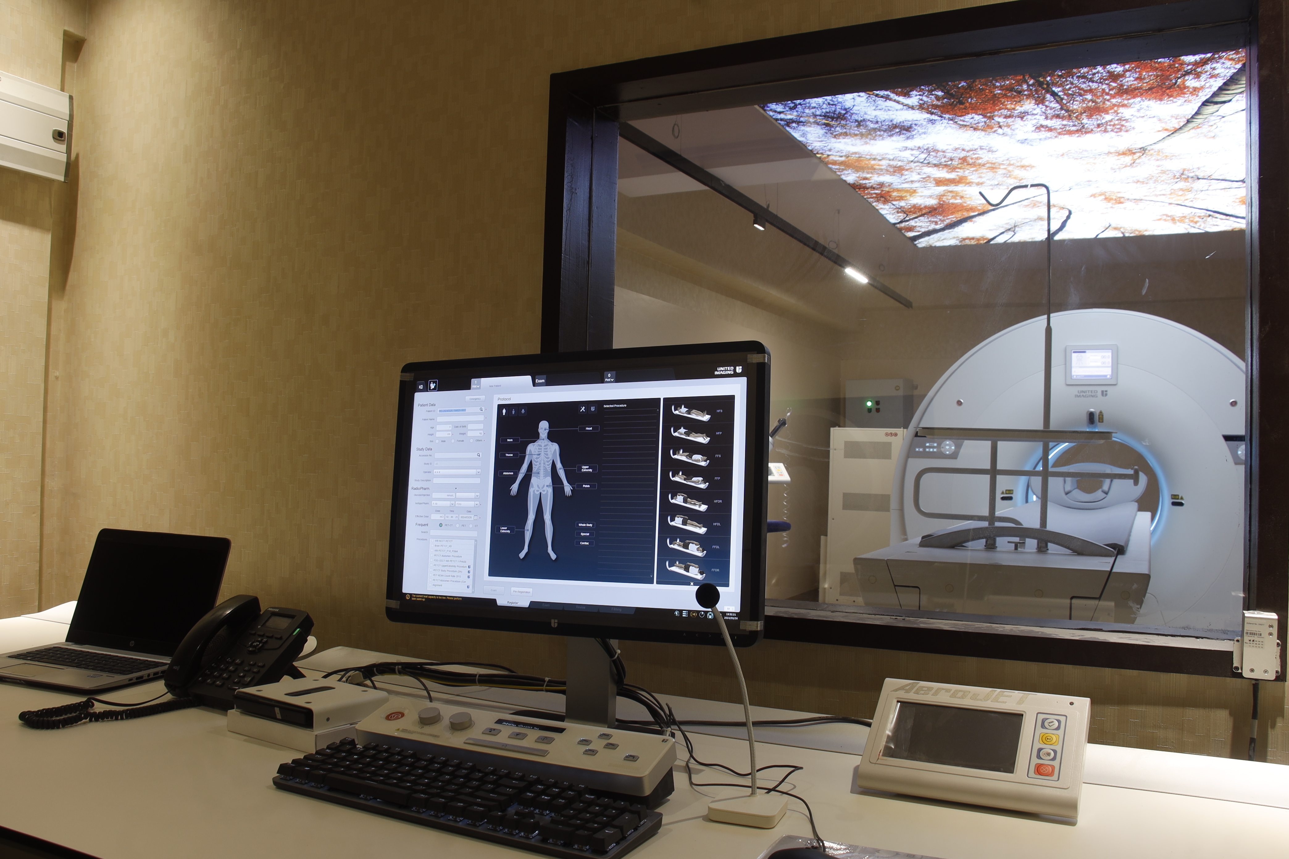
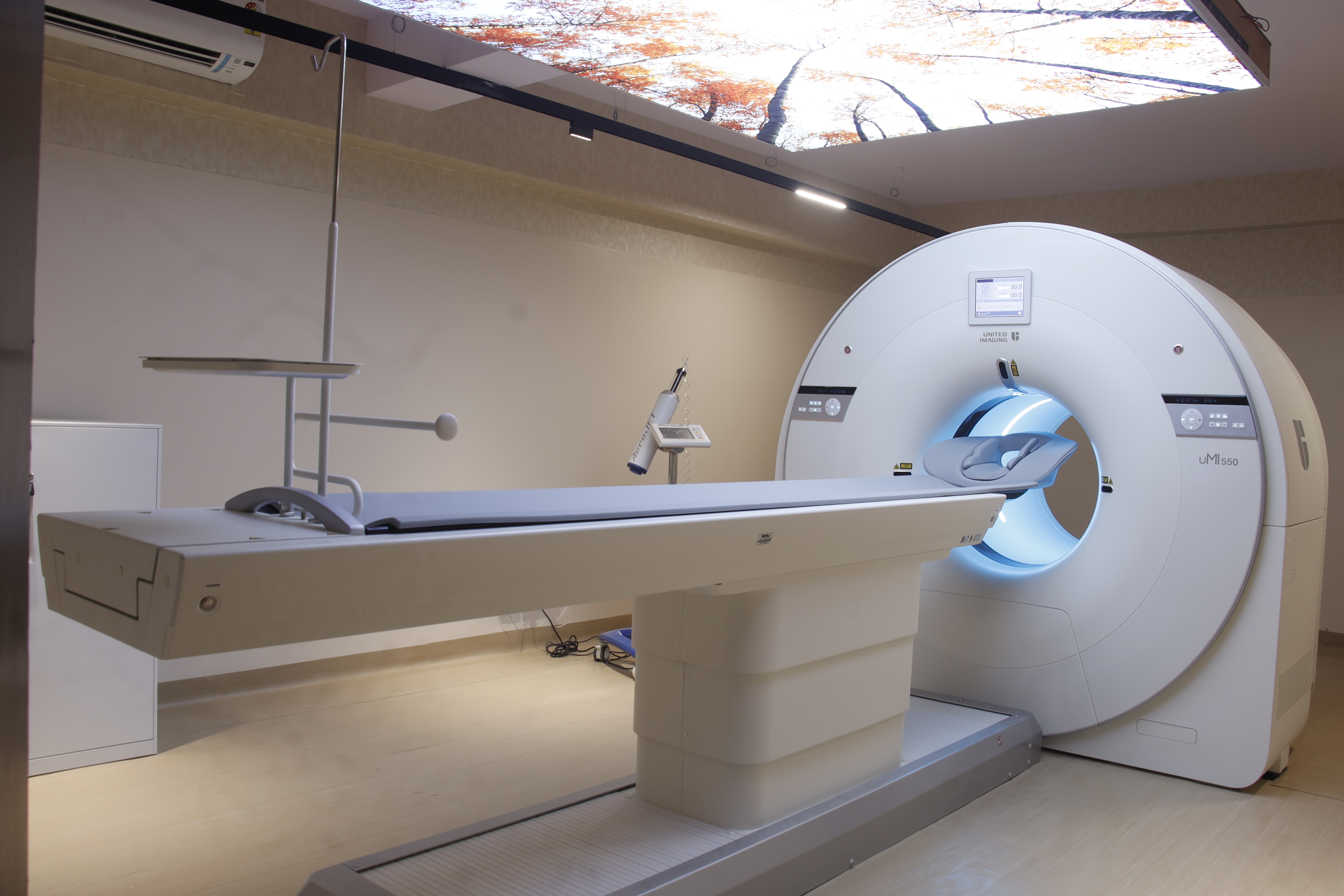
Positron emission tomography–computed tomography (PET-CT) is a nuclear medicine technique which combines, in a single gantry, a positron emission tomography scanner and an x-ray computed tomography scanner, to acquire sequential images from both devices in the same session, which are combined into a single superposed image.
Principle of PET CT (How PET-CT works)
Tumour cells take up glucose under aerobic conditions constitutes the basis for the detection and staging of human cancers with 18F-fluorodeoxyglucose (FDG) and positron emission tomography (PET). 18F-FDG is a structural analogue of 2-deoxyglucose and is used as a tracer of glucose metabolism.Positron Emission Tomography (PET) detects and measures glucose metabolism within the body. In cancers and certain diseases of the heart and brain, the metabolism of glucose may be abnormal.
What is Digital PET-CT
Digital PET-CT imaging is the latest advancement in PET- CT (providing high definition PET images and offering higher sensitivity and higher diagnostic performance). Digital PET-CT imaging is transforming cancer diagnostics with precision, detecting even the smallest lesions missed by conventional imaging techniques, including the analogue PET CT, leading to personalized treatment options and improved patient outcomes. Smallest lesion detectability can change the patient management and improving outcomes in oncological and non-oncological diseases. AI Based reconstruction gives best image quality in lesser injected dose leading to significantly less radiation burden to the patient.
Advantages of digital PET-CT
- Better - image quality, Industries’ best clinical reolution of 1.4 mm.
- Clearer - Crystal clear image quality, Sharper, superior images for accurate disease diagnosis and staging
- Faster - Whole body scanning in just 6-8 min vs. 15-20 min
- Safer - Significantly less radiation to the patient, Digital technology, AI Based reconstruction, Digital Motion correction & Digital Self gating feature avoid repetition of scan leading to less radiation to the patient
- Comfortable - Accommodates patients of all sizes (Upto 250 kg body weight), Compact Gantry Width , Unique in-bore light feature leading to less claustrophobia
- Inclusive - Beyond cancer, digital PET-CT proves valuable in neurology and cardiology, offering precise diagnosis, staging, and treatment evaluation, particularly in early-stage cancer, where tumor size matters most.
Advantages of PET-CT over other imaging modalities
- EARLY detection of disease before appearance of anatomical changes.
- ACCURATE staging and restating of cancers.
- SIMULTANEOUS contrast enhanced CT with metabolic information - cost effective way of imaging the whole body in one sitting.
- QUANTIFICATION of tumor metabolism (SUV) before and after treatment.
- Metabolic GRADING of tumors possible - Impacts treatment decisions.
- SAFE for use in patients with chronic kidney disease, metallic implants, pacemakers, etc.
Test details
Whole body FDG PET-CT Scan
Oncology:
To diagnose primary malignancy : early detection (Because PET images biochemical activity, it can accurately characterize a tumor as benign or malignant, thereby avoiding surgical biopsy when the PET scan is negative)
Initial staging of known malignancy : PET-CT is extremely sensitive in determining the full extent of disease, because of whlie body combined functional and anatomical imaging. Confirmation of metastatic disease allows the physician and patient to more accurately decide how to proceed with the patient's management.
Treatment response evaluation : The level of tumor metabliism (SUV) is compared on PET scans taken before and after a chemotherapy cycle. A successful response seen on a PET scan frequently precedes alterations in anatomy and would therefore be an earlier indicator of tumor response than that seen with other diagnostic modalities.
Post treatment restaging : Once the desired treatment is completed, PET scan is done to check to find any residual cancer cells in the body, if found immediate second line treatment is started.
Follow up to check recurrences : PET is considered to be the most accurate diagnostic procedure to differentiate tumor recurrences from radiation necrosis or post-surgical changes.
Infections:
In PUO - to localise site of occult infection / inflammation.
Tuberculosis - to assess multi focal involvement and response to treatment
Infected Implants/ grafts
Osteomyelitis
Sarcoidosis
Vasculitis
Irritable bowel disease/ syndrome (IBS)
F18 - DOPA PET-CT Scan
Will help to differentiate tremors of parkinson Vs non parkinson disorder - help in deciding line of management.
Dementia
Neuroendocrine tumours
Epilepsy
Brain tumours
FDG Brain PET Scan
Dementia - PET will help to differentiate different types of dementias - Alzheimer, Fronto temporal, senile dementia - helps in decision making on line of management of dementia
Seizure- To look for epileptic focus
Brain tumor recurrence vs radiation necrosis, differentiate benign to malignant tumour.
FDG PET-CT with Triple phase CT
Hepatocellular carcinoma - in cases of raised alphafetoproein with suspicious USG/ CT imaging for HCC.
For better diagnosis of liver, adrenal and pancreatic lesions
F18 PSMA PET-CT Scan
PSMA PET-CT is indicated
(1) in men with newly diagnosed prostate cancer who are at risk for metastatic disease, and
(2) men who have previously been treated for their prostate cancer with curative intent (e.g. with surgery and/or radiation) and now have suspected persistent or recurrent disease based on a rising prostate specific antigen (PSA) level in their blood.
F18-NOTANOC PET-CT Scan
F-18 NOTANOC PET-CT scan is a whole-body examination which gives information regarding the site and extent of primary NET and distant metastasis.
A. Common NETs
(i) Pheochromocytoma
(ii) Medullary Ca thyroid
(iii) Insulinoma
B. Other NETs
Carcinoid, neuroblastoma, Gastrinoma, Merkel Cell Carcinoma, Gatric and Large bowel NET, Somatostatinoma, Pancreatic polypeptidoma, Paraganglioma, Gluganoma, Lung NET, MEN, NET of Pitutary etc.
FDG Cardiac PET Scan
Myocardial viability assessment, to detect hibernating myocardium in post MI patients- help plan treatment protocol - revasularise / medical management
Cardiac sarcoidosis, Myocarditis and infective endocarditis
F18-Choline PET-CT Scan
Most sensitive investigation to localise parathyroid adenoma in case of Primary hyperparathyroidism
More Sensitive than MIBI Parathyroid scan
Better localization of lesion/ multiple lesions
Prior appointment is mandatory/ preferable.
One attendant is desirable.
Patients are requested to report for their scan, 30 minutes before their appointment time.
Pregnancy is a contra-indication. Please inform if there is any chance of pregnancy or history of missed period. Please inform if the patient is a nursing mother (breast-feeding).
Overnight or minimum 4 - 6 hours fasting is required prior to appointment time (except diabetic, diabetic patients please read special instructions below). Drinking of water is allowed but no other liquid. No need to hold urine.
Parentral feeding (IV dextrose drip) should also be withheld for 4-6 hours (Tube feeding).
Intravenous fluid containing dextrose should be withheld for 12 hours (for admitted patients).
Patients are requested to remove all the ornaments before coming for the scan.
PET-CT scans are completely painless.
Dress comfortably and warmly, as scanner rooms are cool.
If you need pain medication, please bring it with you.
Patients should avoid alcohol, sweets and strenuous exercise at least two days prior to the PET-CT study.
Pregnant women / children should not accompany the patient
Patients are instructed to bring their Recent Blood sugar, Blood Urea and S. Creatinine reports – maximum one month old, if not done than these tests can be done here on the same day prior to the PET-CT scan.
Patients are instructed to bring all their previous investigations reports and films (MRI and CT reports, histopathogy reports), discharge summaries and doctor’s prescription. Please must carry previous PET-CT (CD/ DICOM images) with report and doctor’s prescription for PET-CT on the day of test.
Diabetic patients should inform us about their blood sugar report in advance.
Blood glucose level should ideally be less than 200mg/dl, if it is more then we might have to delay or postponed the test.
Patients should take a light breakfast along with their anti-diabetic medicines between 5- 6 am or 4-6 hours prior to the scheduled time and come for scan at least 2 hours before the scheduled time.
Patients should bring all the diabetic medicines along with him / her.
Upon arriving at the PET-CT department, you will receive an IV-injection of radioactive glucose like FDG/ PSMA, following which you shall rest for approximately 30-90 minutes in isolation to allow FDG/ PSMA to distribute throughout your body.
Then you will be asked to empty your bladder and change clothes, then lie down on the PET-CT machine.
The scan takes approximately 6-8 minutes, depending upon the type of scan you are undergoing. It is important that you lie still during this process.
It is not noisy and there is no tunnel-like feeling as in an MRI.
The gantry is well lit.
You should plan on being at the center for approximately 2-3 hours. In case a delayed scan is needed, then there may be additional stay.
Once the PET scan is complete, you will be able to leave the center. Make sure you drink plenty of water or other fluids throughout the day to flush the FDG from the body, unless regulated by your doctor.
You should avoid close contact with other people, especially children below 5 years of age and pregnant females during the next 24 hours.
A senior nuclear medicine physician and a radiologist interpret the PET-CT scan, and the results will be ready within 24-48 hours depending upon the test. You should contact your referring doctor to discuss the results.
For your PET scan, a radioactive drug called a tracer will be injected into a vein. CT scan also uses some radiation from x-rays. Because the amount of radiation you're exposed to in the tracer is small, the risk of negative effects from the radiation is low. But the tracer might expose your unborn baby to radiation if you are pregnant, so please inform the technologist/ doctor if there is any chance of pregnancy.
Our's Vs. Other's
| Digital PET | Analogue PET |
|---|---|
| Latest advancement in PET-CT (provide high definition PET images and offer higher sensitivity and higher diagnostic performance) | Existing most PET-CT machines |
| Highest Resolution -- 1.4 mm (Smallest lesion detectability) | Resolution > 2 mm |
| Smallest lesion detectability can change the patient management and improving outcomes in oncological and non-oncological diseases | Can miss small lesions which can affect Staging of disease |
| Shorter scan time 8 min (increases the comfort level of elderly, children & sick patients) | Longer scan time 15-20 min |
| Less radiation burden to the patient | More radiation burden to the patient |
| Al Based reconstruction gives best image quality in lesser injected dose (Crystal Clear Image Quality) | Not so sharp images |
| Reporting by Senior Nuclear Medicine Physician as well as radiologist for better diagnosis (Experience of more than 20 years) | Only reported by Nuclear Medicine Physicain |
| All investigations under one roof- MRI, CT, USG, Path Lab, Dexa, Mammography, Fibroscan, Echo, Stress Echo, ECG, TMT, EPS, CBCT | Refer to other centres for other tests |
| Wide Gantry (70 cms) Accommodates larger patients | Smaller Gantry |
| Compact Gantry Width reduces the Claustrophobia. | Heavy Gantry - more Claustrophobia |
| Unique in-bore light feature - less claustrophobia | No in bore light - more Claustrophobia |
| Highest Weight wearing capacity of table i.e. 250 KG | Weight wearing capacity of table - Not suitable for overweight patients |
| Digital Motion correction & Digital Self gating feature avoid repetition of scan | No such features - Repeat the scan - increases radiation burden to patient |
Whole Body FDG
-

Home Collection: Not Available
-

Report Available: 1 Day
Whole Body FDG with Triple Phase CT
-
Home Collection: Not Available
-
Report Available: 1 Day
Extra CD Making Charges (@ 500 Rs).
-
Home Collection: Not Available
-
Report Available: 1 Day
EXTRA IMAGES CHARGES (@300Rs per film).
-
Home Collection: Not Available
-
Report Available: 1 Day
F18-Choline
-
Home Collection: Not Available
-
Report Available: 1 Day
F18-DOPA
-
Home Collection: Not Available
-
Report Available: 1 Day
F18-Notanoc
-
Home Collection: Not Available
-
Report Available: 1 Day
F18-PSMA
-
Home Collection: Not Available
-
Report Available: 1 Day
FDG Brain
-
Home Collection: Not Available
-
Report Available: 1 Day
FDG-Cardiac
-
Home Collection: Not Available
-
Report Available: 1 Day
Have a query or suggestion for us? Feel free to drop us a message and we'll get back to you as soon as possible.